

OIC orthopaedic surgical team in operating room — high-value implant solutions
Medical team — OIC value-based orthopaedic implants for hospitals and surgeons
Active patients running — OIC orthopaedic implants enable recovery and return to life
News & Perspectives

Orthopaedic Insights & Industry Blog

Exploring Value-Based Healthcare & Clinical Progress

0%

Ankle Syndesmotic Repair Systems have become increasingly important as surgeons seek stable, anatomically accurate fixation for distal tibiofibular injuries. Orthopedic trauma reports from MarketsandMarkets and Grand View Research identify sustained demand for fixation technologies, driven by sports injuries, ankle fractures, and minimally invasive procedures. However, broad market growth does not prove that one repair system is clinically superior.

The meaningful comparison begins in the operating room. Surgeons assess reduction accuracy, fixation strength, implant flexibility, drilling requirements, and postoperative rehabilitation. Common options include trans-syndesmotic screws, suture-button constructs, and hybrid systems. Each design leaves a different footprint in bone and soft tissue. Small details matter, including tunnel angle, cortical purchase, and the risk of losing reduction during weight-bearing.

Michel P. J. van den Bekerom and colleagues stated, “The gold standard for the diagnosis of syndesmotic instability is still unclear.” That caution remains relevant when evaluating modern Ankle Syndesmotic Repair Systems. A device may appear technically elegant, yet patient anatomy and injury pattern can change the result. Peer-reviewed reviews in Injury and the Journal of Orthopaedic Trauma also emphasize accurate reduction as a major predictor of outcome. This article compares leading systems through clinical evidence, surgeon experience, design characteristics, and reported complications. The ranking is not absolute. Evidence quality varies, and some commercial claims still require stronger independent validation.
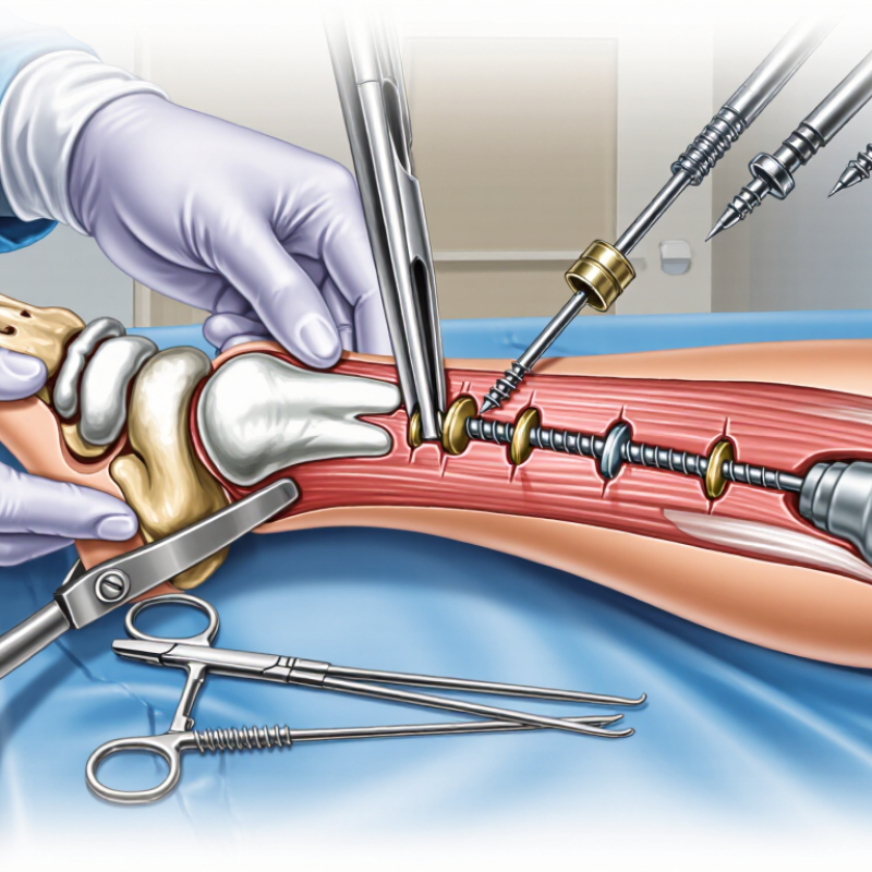
What Are the Top Ankle Syndesmotic Repair Systems?

Ankle Syndesmosis: Anatomy, Function, and Common Injuries

The ankle syndesmosis is a compact ligament complex between the tibia and fibula. It includes the anterior and posterior tibiofibular ligaments, the interosseous ligament, and its connecting membrane. Together, these tissues maintain the ankle mortise, the shallow socket surrounding the talus. During walking, they allow tiny movements while resisting excessive separation. That balance is easy to underestimate.

Syndesmotic injuries often occur when the foot twists outward while the leg rotates inward. They may accompany ankle fractures, but some appear without a broken bone. Pain above the ankle is common. Swelling can be mild, and bruising may develop later. Clinicians assess tenderness, stability, walking tolerance, and rotation pain. Weight-bearing radiographs can help, while stress views or advanced imaging may clarify uncertain cases. Examination is not perfect. Early symptoms can mislead even experienced practitioners.

When the syndesmosis remains stable, protected weight bearing and rehabilitation may be appropriate. Unstable injuries may require fixation after the bone position is restored. Common repair systems include rigid trans-syndesmotic screws, flexible suture-button constructs, and combined fixation methods. Screws provide firm stabilization but may restrict natural motion or require later removal. Flexible constructs can permit controlled movement, although positioning and bone quality still matter. The best choice depends on injury pattern, fracture repair, tissue condition, and surgeon judgment. No system works equally well for every ankle. Careful reduction is crucial; even a small malalignment can change joint pressure and prolong recovery.

When Syndesmotic Repair Is Needed

When syndesmotic repair is needed, the key question is instability, not simply the presence of a fracture. The syndesmosis connects the tibia and fibula above the ankle joint. If it widens during stress testing, healing may leave the ankle painful and mechanically uneven.

The British Orthopaedic Association’s BOAST Ankle Fracture guidance recommends assessing syndesmotic stability after fracture fixation. Surgeons commonly use a trans-syndesmotic screw, a flexible suture-button construct, or a hybrid system. A 2022 systematic review in Foot & Ankle Orthopaedics found that flexible fixation generally produced comparable functional outcomes to screw fixation. It also reported fewer routine implant-removal procedures. Evidence remains mixed.

Clinical experience matters here. A high ankle sprain can hide behind swelling, bruising, and a seemingly acceptable X-ray. Weight-bearing radiographs, computed tomography, and intraoperative stress tests can reveal different details. The American College of Radiology rates CT as useful for evaluating complex ankle fractures and syndesmotic alignment.

A small measurement can change the plan. Yet no single distance should dictate treatment. Patient age, bone quality, fracture pattern, activity level, and surgical timing also matter. I would be cautious about calling every subtle widening unstable. That judgment still requires trained examination and imaging review.

Main Types of Ankle Syndesmotic Repair Systems

Ankle syndesmotic repair systems generally fall into three main types: rigid screws, flexible suture-button devices, and hybrid constructs. Syndesmotic screws remain familiar because they provide strong initial fixation across the tibia and fibula. Their limitations are important. Screw breakage, loosening, and routine removal can lead to another procedure. A 2021 meta-analysis of comparative clinical studies found that screw fixation often required more implant removal than flexible fixation.

Suture-button systems use strong fibers and small fixation buttons to maintain reduction while allowing limited physiologic movement. This design may reduce routine removal and support earlier functional recovery. A 2023 systematic review in Foot & Ankle Orthopaedics reported lower reoperation rates with flexible fixation in several comparative studies, although study quality varied. Results are not perfectly consistent. Patient activity, ligament injury pattern, and reduction accuracy still matter greatly.

Hybrid fixation combines a screw with a flexible device. Surgeons may consider it for high-energy injuries, poor bone quality, or unstable fractures. It can offer extra resistance, but it also adds implants, cost, and technical demands. The American Orthopaedic Foot & Ankle Society emphasizes imaging and clinical assessment when evaluating syndesmotic stability. In practice, the best system is not chosen from a catalog alone. Intraoperative testing remains essential. Even experienced teams can miss subtle malreduction, and that problem deserves more attention.

What Are the Top Ankle Syndesmotic Repair Systems? – Main Types of Ankle Syndesmotic Repair Systems
Repair System Type Core Construct Typical Indications Biomechanical Characteristics Key Advantages Main Limitations and Risks Common Postoperative Considerations
Trans-syndesmotic Screw Fixation One or more rigid screws pass from the fibula into the tibia, usually through a fibular plate or directly through the fibula.
Rigid fixationMetal screw
Acute syndesmotic instability associated with ankle fractures, especially when the syndesmosis remains unstable after fibular and medial-column reduction. Provides strong resistance to syndesmotic separation but restricts normal physiologic motion between the distal tibia and fibula. Familiar surgical technique; widely available; provides predictable initial fixation; useful when substantial stability is required. Screw loosening, breakage, malreduction, loss of reduction, and irritation may occur. Routine removal is not universally required and depends on symptoms, healing, and local practice. Weight-bearing progression is often more cautious than with flexible fixation. Follow-up commonly includes clinical assessment and radiographs to evaluate reduction and hardware integrity.
Suture-Button Fixation A high-strength flexible suture connects two cortical buttons positioned on the fibular and tibial sides of the syndesmosis.
Dynamic fixationFlexible construct
Acute isolated syndesmotic instability or syndesmotic disruption accompanying an ankle fracture when adequate reduction can be achieved and maintained. Allows limited physiologic micromotion while resisting excessive widening of the distal tibiofibular relationship. Usually avoids planned hardware removal; may permit earlier functional rehabilitation; less likely than a rigid screw to break from repetitive physiologic motion. Malpositioned tunnels, button migration, local irritation, infection, osteolysis, and recurrent widening are possible. It requires accurate reduction and careful tunnel placement. Rehabilitation is guided by fracture healing, soft-tissue condition, fixation security, and surgeon protocol rather than by the device alone. Imaging is used to confirm maintained alignment.
Hybrid Screw–Suture-Button Fixation Combines a rigid trans-syndesmotic screw with a flexible suture-button construct, often with the screw providing initial compression and the flexible device supporting dynamic stability.
Combined fixationRigid + dynamic
High-grade or multidirectional syndesmotic instability, complex ankle fractures, or cases in which additional resistance to rotational and translational forces is considered necessary. Combines high initial stiffness with controlled motion, although the optimal combination and positioning depend on the injury pattern. May provide enhanced resistance to diastasis and rotation compared with either method alone in selected unstable injuries. More implants and greater surgical complexity; increased risk of malposition, soft-tissue irritation, and implant-related symptoms. Screw removal may still be considered if symptomatic. Rehabilitation and weight-bearing decisions are usually individualized. Radiographic monitoring is important because both rigid and flexible components must remain well positioned.
Plate-Assisted Syndesmotic Fixation A fibular plate stabilizes the distal fibula and serves as a platform for one or more syndesmotic screws or flexible fixation devices.
Fracture fixationAnatomic support
Syndesmotic disruption with an unstable fibular fracture, comminution, poor bone quality, or a need to restore fibular length and rotation before syndesmotic stabilization. The plate restores and maintains fibular alignment, while the attached screw or flexible device controls tibiofibular separation. Addresses both fibular fracture stability and syndesmotic instability; may improve control of fibular length, rotation, and alignment. Plate prominence, soft-tissue irritation, infection, peroneal tendon irritation, and possible secondary surgery may occur. Fixation cannot compensate for inaccurate fracture or syndesmotic reduction. The fracture and syndesmosis are assessed together during follow-up. Hardware removal is considered only when clinically indicated or required by symptoms and healing status.
Multiple-Screw or Multi-Point Fixation Two or more trans-syndesmotic screws, or fixation at more than one level, are used to increase resistance to separation and rotation.
High-stability constructMultiple fixation points
Severe syndesmotic disruption, proximal extension of the injury, marked instability, osteoporotic bone, or selected revision and salvage situations. Increases construct stiffness and load distribution but may further limit physiologic distal tibiofibular motion. May improve fixation strength in highly unstable patterns and compromised bone, when appropriately planned. Greater implant burden; higher potential for malreduction, screw breakage, stiffness, and soft-tissue irritation. The additional fixation does not eliminate the need for accurate reduction. A longer protection period may be selected for complex injuries. Serial clinical and radiographic evaluation is commonly used to monitor reduction, healing, and hardware-related symptoms.
Syndesmotic Fixation with Supplemental Medial-Side Stabilization A primary syndesmotic device is combined with repair or fixation of associated medial structures, such as the deltoid ligament or medial malleolus, when clinically indicated.
Combined injury managementMulticolumn stability
Rotationally unstable ankle injuries with medial-column disruption, medial malleolar fracture, or persistent medial instability after fixation of the lateral and syndesmotic components. Treats the syndesmosis as part of the complete ankle stability system rather than as an isolated structure. Can improve overall ankle stability when medial and syndesmotic injuries coexist; supports restoration of the ankle mortise when indicated. Requires accurate diagnosis of all injured structures and may involve additional dissection, implants, operative time, and rehabilitation restrictions. The rehabilitation plan is based on the most restrictive component of the injury. Assessment should include mortise alignment, medial stability, syndesmotic reduction, and fracture healing.
Clinical note: The most appropriate ankle syndesmotic repair system depends on the injury pattern, quality of reduction, associated fractures or ligament injuries, bone quality, patient activity level, and surgeon experience. No single construct is universally superior, and accurate anatomic reduction remains a central determinant of outcome.

How Leading Repair Systems Differ

Leading ankle syndesmotic repair systems differ mainly in fixation method, adjustability, and surgical handling. Some use a fixed screw, while others use flexible suture-based constructs. Adjustable loops can help surgeons fine-tune compression during reduction. However, flexibility does not automatically mean better stability.

A screw may provide firm fixation, especially when bone quality is reliable. It can also require later removal or monitoring for breakage. Suture-based systems may reduce implant prominence and support earlier ankle movement.

Their performance depends heavily on accurate reduction and secure cortical purchase. Small placement errors can change ankle mechanics. That detail is easy to underestimate.

The best system depends on the injury pattern, fracture level, bone quality, and surgeon experience. Imaging before and during surgery remains important. A practical surgeon may also consider instrument simplicity, revision options, and rehabilitation demands. In a crowded operating room, clear tension markers can prevent avoidable mistakes.

No system is perfect. Even strong evidence has limits, because studies often compare different techniques and patient groups. Careful follow-up matters, especially when pain, widening, or restricted motion continues.

In my view, the most reliable choice is not always the newest design. It is the system that allows accurate reduction, stable fixation, and consistent postoperative assessment.

Surgical Steps, Recovery, and Potential Complications

What Are the Top Ankle Syndesmotic Repair Systems?

Surgical Steps, Recovery, and Potential Complications

Ankle syndesmotic repair stabilizes the ligaments between the tibia and fibula. Common systems include rigid screws, flexible suture-button devices, and hybrid constructs. The best choice depends on injury severity, fracture pattern, bone quality, and the surgeon’s assessment of stability.

During surgery, the team first restores the ankle joint and confirms alignment with imaging. The surgeon then reduces the fibula and tests the syndesmosis under stress. A fixation device is placed across the ankle, while the position is checked from multiple angles. Accurate reduction matters. Even a small rotational error can affect walking mechanics and long-term comfort.

Recovery usually begins with a splint, elevation, and limited weight-bearing. Some patients avoid weight for several weeks, depending on fractures and fixation strength. Gentle motion may start early, but loading progresses gradually through physical therapy. Strength, balance, and calf control require patience. Real recovery is messier than diagrams suggest.

Potential complications include infection, stiffness, nerve irritation, malreduction, and loss of fixation. Rigid screws may loosen or break, and removal is sometimes considered after healing. Flexible systems can also fail if the ankle remains unstable or the fixation tunnel is poorly positioned. Persistent pain deserves review, not dismissal. Follow-up imaging helps identify problems before they become harder to correct.

What Are the Top Ankle Syndesmotic Repair Systems?

Typical postoperative timing by fixation construct

Syndesmotic screw fixation commonly uses protected weight-bearing for approximately 6–8 weeks, while suture-button constructs may permit earlier progression in selected patients. Hybrid fixation generally follows a more conservative 6–8 week protected-weight-bearing protocol.

These are representative clinical protocol windows rather than guaranteed outcomes. Timing depends on fracture stability, ligament healing, bone quality, associated injuries, imaging, and the surgeon’s rehabilitation plan. Potential complications include malreduction, recurrent syndesmotic instability, implant loosening or breakage, infection, wound problems, nerve irritation, stiffness, and post-traumatic ankle arthritis.

Values represent commonly reported clinical ranges; individual treatment plans may differ.

FAQS

What is the ankle syndesmosis?

It is a compact ligament complex between the tibia and fibula. It helps maintain the ankle socket around the talus. Tiny movements occur during walking. Stability still matters.

What tissues make up the syndesmosis?

It includes the anterior and posterior tibiofibular ligaments. It also contains the interosseous ligament and connecting membrane. Together, they resist excessive separation between the lower-leg bones.

How do syndesmotic injuries usually happen?

They often occur when the foot twists outward while the leg rotates inward. This force can stretch or tear the ligaments. A fracture may occur, but not always.

What symptoms can suggest a syndesmotic injury?

Pain above the ankle is common. Swelling may be mild. Bruising can appear later. Rotation pain and difficulty bearing weight may also occur.

How is this injury evaluated?

A clinician checks tenderness, ankle stability, walking tolerance, and pain during rotation. Weight-bearing X-rays can help. Stress views or advanced imaging may clarify uncertain cases.

Can an examination miss the injury?

Yes. Early symptoms can mislead, even for experienced clinicians. Imaging and follow-up may reveal problems later. That uncertainty deserves respect.

How are stable syndesmotic injuries commonly managed?

Stable injuries may receive protected weight bearing and rehabilitation. Recovery usually involves gradual movement and strengthening. The exact plan depends on tissue damage and clinical findings.

When might fixation be considered?

Unstable injuries may need fixation after the bones are correctly restored. Options include rigid screws, flexible suture-based constructs, or combined methods. Accurate reduction remains essential.

How do rigid screws and flexible constructs differ?

Screws provide firm stabilization but may restrict natural motion. They might require later removal or monitoring for breakage. Flexible constructs permit controlled movement, though placement must be precise.

What factors influence the repair method?

The injury pattern, fracture level, bone quality, tissue condition, and surgeon experience all matter. Instrument simplicity and rehabilitation needs may also influence the decision. Newer is not automatically better.

Conclusion

Ankle Syndesmotic Repair Systems are designed to stabilize the ligaments that connect the lower ends of the tibia and fibula, helping restore normal ankle alignment after a severe sprain, fracture, or syndesmotic disruption. This injury may cause pain, swelling, instability, and difficulty bearing weight. Repair is generally considered when the joint is unstable, the ligaments are significantly separated, or conservative treatment is unlikely to provide reliable healing.

Common system types include flexible fixation devices, screw-based fixation, and hybrid approaches. They differ in flexibility, fixation strength, adjustment options, and whether a later procedure may be needed for removal. During surgery, the joint is carefully realigned, the fixation is positioned across the syndesmosis, and stability is checked before closure. Recovery usually involves a period of protection, gradual weight bearing, and physical therapy to restore motion, strength, and balance. Potential complications include infection, nerve irritation, malalignment, hardware problems, restricted movement, or recurrent instability, so treatment should be individualized through careful clinical and imaging assessment.

Oliver

Oliver

Oliver is a dedicated marketing professional with a deep expertise in our company's products and services. With a keen understanding of industry trends and consumer behavior, he crafts insightful blog posts that not only educate our audience but also highlight the unique benefits of our offerings.......